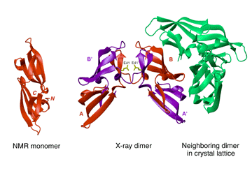
Bewley, C.A., Gustafson, K.R., Boyd, M.R., Covell, D.G., Bax, A., Clore, G.M. & Gronenborn, A.M. (1998) Solution structure of cyanovirin-N, a potent HIV-inactivating protein. Nature Struct. Biol. 5, 571-578. pubmed PDF
Yang, F., Bewley, C.A., Louis, J.M., Gustafson, K.R., Boyd, M.R., Gronenborn, A.M., Clore, G.M. & Wlodawer, A. (1999) Crystal structure of cyanovirin-N, a potent HIV-inactivating protein, shows unexpected domain swapping. J. Mol. Biol. 288, 403-412. pubmed PDF
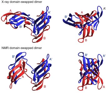
Bewley, C.A. & Clore, G.M. (2000) Determination of the relative orientation of the two halves of the domain-swapped dimer of cyanovirin-N in solution using dipolar couplings and rigid body minimization. J. Am. Chem. Soc. 122, 6009-6016. PDF
Clore, G.M. & Bewley, C.A. (2002) Using conjoined rigid body / torsion angle simulated annealing to determine the relative orientation of covalently linked protein domains from dipolar couplings. J. Magn. Reson. 154, 329-335. pubmed PDF
|
Disclaimer: Dr. Clore created this website in his personal capacity. The information on this site and views expressed are his own and do not necessarily represent the views of the National Institutes of Health or the United States Government
|